
µPET/CT Albira
The current configuration of µPET/CT scanner Albira (manufacturer Carestream Health, Inc., now Bruker Corp.) consists of two systems – see Fig. 1 (overall view) and Fig. 2 (function modules): a system for positron emission tomography (PET) with one ring of detectors with high resolution and a system for computer tomography (CT). The systems can be used either isolated or in combination for fusion imaging of physiological/functional processes (PET) together with detailed imaging of anatomical structures (CT) of small laboratory animals. Furthermore, the scanner is equipped with monitoring system Biopac (MP150, manufacturer Biopac Systems, Inc.) for continuous monitoring of basic physiological functions and parameters of animals under anaesthesia during scanning (EKG-heart rate, blood pressure, respiration, oxygen consumption, blood gases, etc.).

Fig. 1: μPET/CT scanner Albira – overall view
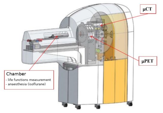
Fig. 2: μPET/CT scanner Albira – function modules
PET module consists of one ring with 8 detectors composed of large (50×50 mm) and 10 mm thick continuum lutetium yttrium orthosilicate (LYSO)-based scanners. The scanners are in direct contact with the position sensitive photomultiplier tubes (PSPMT) for detection of light flashes and with patent-protected dedicated electronics. The system enables 3D imaging including determination of the interaction depth based on shape analysis of detected light signal with lower than 1.3 mm resolution. Longitudinal and transverse field of vision of the ring of detectors (axial field of view FOV and transaxial FOV) is 40 mm and 80 mm.
The Albira CT module consists of the X-ray source, X-ray tube for industry applications with tuneable voltage between 4 kV and 50 kV and tuneable current between 0 and 1 mA and corresponding X-ray detector, which consists of CsI monocrystal and photodiodes. The nominal X-ray focal spot size of 35 mm is suitable for X-ray imaging of small laboratory animals; the axial × transaxial FOV is 70 × 70 mm. The X-ray tube and detector are mounted to a board and located against each other in the angle of 180°. The board is rotational enabling 360° rotation with precise position setting better than 0.01°. The detector crystal provides high resolution (50 mm) on a big area of 120 × 120 mm, therefore enabling whole mouse body imaging in one 360° turn. The detector area is composed of 2 400 × 2 400 pixels and the minimal achievable resolution of the CT system is 90 mm.
The µPET/CT scanner operating software Albira Software Suite v. 09-0117 was installed in 2015 and compose of four fundamental application modules: Albira Manager, Albira Acquirer, Albira Reconstructor and Albira Supervisor. Evaluation and quantification PET and CT scans is done using licensed software PMOD v. 3.403 (PMOD Technologies, Ltd., Zürich, Switzerland), which was developed for advanced PET image reconstruction in preclinical research. The Albira scanner is operated by PC with the evaluating software Carimas v. 2.9, developed in Turku PET Centre, Finland.