
Quantitative measurement of capillaries
We study capillary bed using stereology and 3D image analysis.
The object of our study is an effect of the ionizing radiation on blood capillaries in various tissues and organs. We collaborate with team of Dr. Mao (Department of Radiation Medicine, Loma Linda University, CA, USA) on project focused on proton therapy.
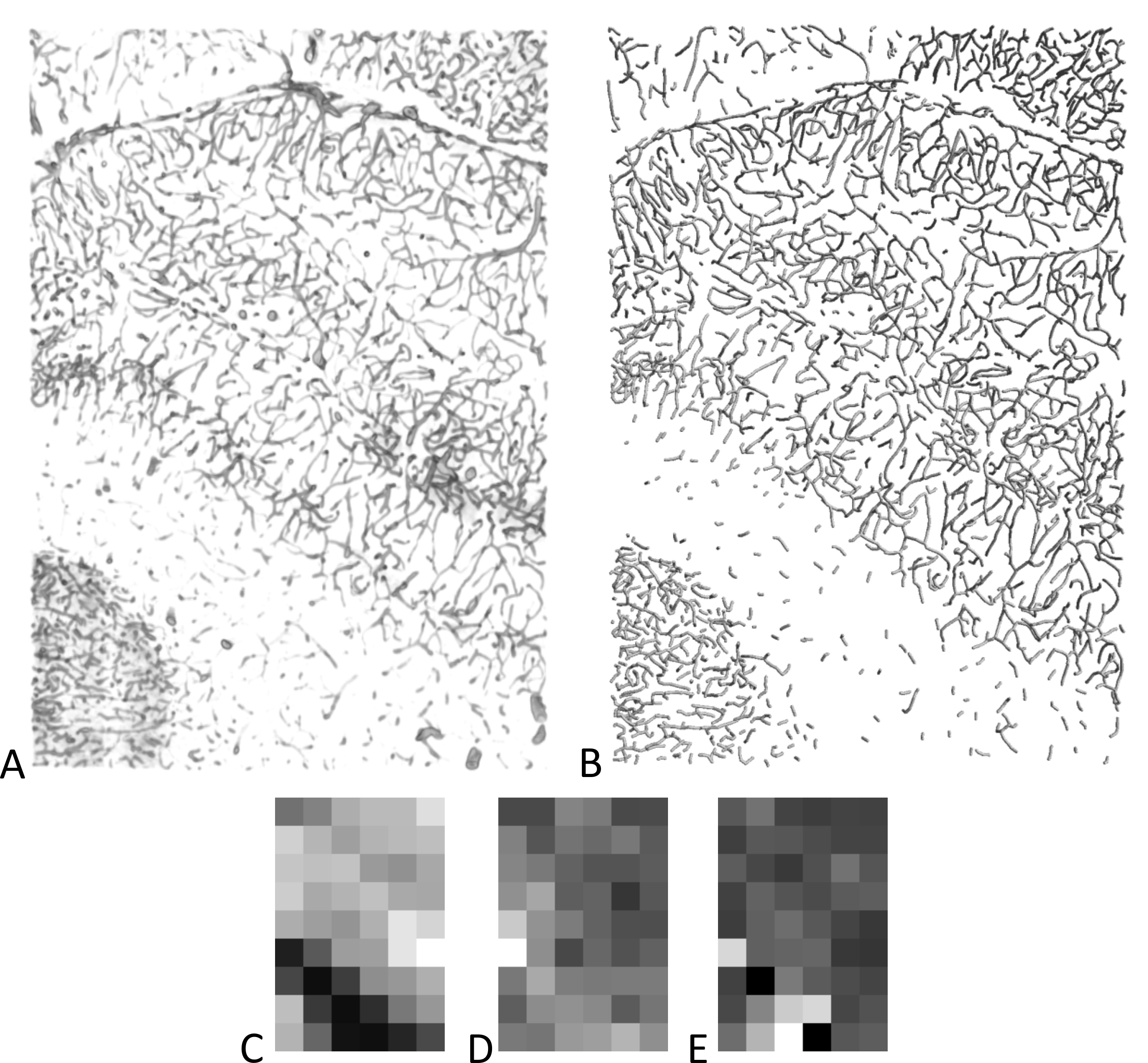
A) Volume rendering of composite images (70 confocal stacks in 10 rows and 7 columns, 1762×2519×80 µm) and B) surface rendering of skeletonized capillaries as cylinders. Images were obtained by a confocal microscope from perfusion stained samples of rat brain. C–E) Local measurements of the data in figure A, B. The image was split into 6×9 tiles before measurement and the value was represented by the gray level. Minimal value is represented by black. C) Length density, maximal value is 695 mm-2, D) anisotropy characteristics (0 to 1), maximal value is 0.74, E) average branch length, maximal value is 0.7 mm.