New diagnostic approaches to mitochondrial diseases
Using lymphocytes isolated from peripheral blood, we try to develop new diagnostic protocols for patients with suspected mitochondrial diseases.
This is a joint cooperative project between Institute of Physiology AS CR, Department of Paediatrics and Adolescent Medicine at 1st Faculty of Medicine, Charles University and Thomayer hospital. Diagnostics of a substantial part of mitochondrial diseases relies on clinical symptoms and biochemical analysis of energetic function and content of individual mitochondrial proteins in patient tissues – mainly in bioptic samples of skeletal muscle and cell cultures of skin fibroblasts. However, both these approaches have significant drawbacks. Muscle biopsy allows for an instant, single biochemical analysis of the mitochondrial quantity and function, but it is often complicated by muscle atrophy caused by mitochondrial disorders. Also, due to its invasive nature, the biopsy is often refused by the parents of the patient, and the same holds true for the skin biopsy, necessary for establishing fibroblast cell culture. While the cell culture allows for more systematic and repeated analysis of mitochondria, the diagnostic results are obtained late, after several weeks or even months. While diagnostics of mtDNA encoded defects in lymphocytes is problematic due to their heteroplasmic nature, they appear to be highly perspective in cases of autosomally-transmitted, homozygous mutations. Here, the analysis of mitochondrial energetics apparatus in lymphocytes, with the help of sensitive functional methods and detection of quantitative and qualitative changes in OXPHOS proteins, could become an attainable alternative method
The aim is to improve and simplify diagnostics of mitochondrial diseases of nuclear genetic origin by analysis of mitochondrial energetics apparatus in peripheral blood lymphocytes, with the help of highly sensitive functional methods, detection of quantitative and qualitative changes in OXPHOS complexes and changes in mitochondrial metabolites.
|
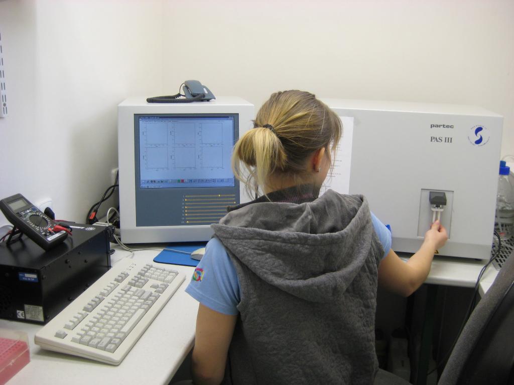
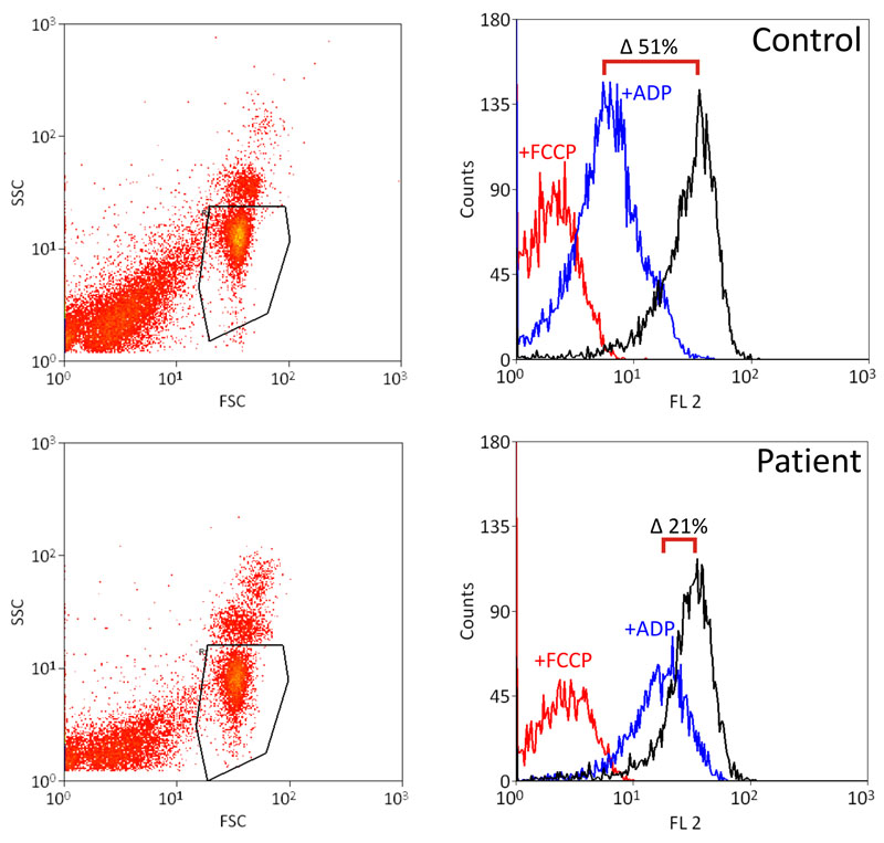
Measurement of mitochondrial membrane potential on flow cytometer. |
And the result - patients with ATP synthase defect show lower discharge of membrane potential after ADP addition. |