
Nanocellulose as a promising cell carrier for applications in regenerative medicine
Cellulose in the form of a fabric has been used for thousands of years as a traditional wound dressing material. Nowadays, it can be used not only for passive wound covering, but also for active wound healing, e.g. with controlled delivery of various drugs or cells for regenerating the damaged tissue. A suitable form of cellulose for this purpose is nanocellulose, i.e. cellulose in the form of nanofibrils, simulating the architecture of the native extracellular matrix. This form of cellulose is produced by some species of bacteria, or can be isolated from higher plants, including wood. In our experiments, we have focused on the development of “intelligent” wound dressings, capable of delivering skin and stem cells into skin wounds. These dressings are based on electrically-charged cellulose nanofibrils, attached to a microfibrous cellulose fabric. Anionic nanocellulose provided a suitable substrate for the adhesion and growth of human dermal fibroblasts and human adipose tissue-derived stem cells, while cationic nanocellulose provided better support for cell-cell adhesion and for the formation of cell aggregates, which was apparent mainly in fibroblasts (Fig. 1). This difference was due to the preferential adsorption of albumin from the serum supplement of the culture medium, which is non-adhesive for cells, on cationic nanocellulose. However, both types of nanocellulose are useful in regenerative medicine. Anionic nanocellulose is suitable for creating continuous cell sheets, which can be delivered into the wound either spontaneously or after release from the substrate using cellulase enzymes, while cationic cellulose is suitable for creating cell spheroids, i.e. important structures for developing organoids and for tissue engineering.
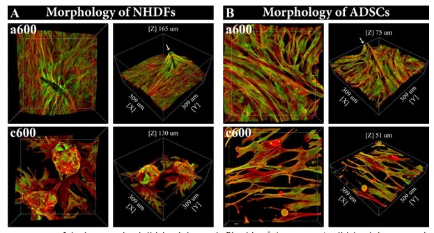
Fig. 1 Morphology of normal human dermal fibroblasts (NHDFs; A) and human adipose tissue-derived stem cells (ADSCs; B), guided by the topography of cellulose meshes coated with anionic or cationic cellulose nanofibrils (a600, c600, respectively) after seven days of cultivation. 3D projection of microscopy images (front view and side view) of the cells on the material. F-actin of the cell cytoskeleton is stained in red, vinculin in the cells is stained in green. Confocal microscope with objective magnification 40x.
Pajorova J, Skogberg A, Hadraba D, Broz A, Travnickova M, Zikmundova M, Honkanen M, Hannula M, Lahtinen P, Tomkova M, Bacakova L, Kallio P. A cellulose mesh with charged nanocellulose coatings as a promising carrier of skin and stem cells for regenerative applications. Biomacromolecules, 2020, 21: 4857-4870, https://dx.doi.org/10.1021/acs.biomac.0c01097; IF = 6.092; DOI